Digital radiography software for industrial NDT
See every indication.
Prove every reading.
Open DICONDE files from any DR system in your browser, compressed or not. Enhance them on the GPU, measure in millimetres, check image quality against ISO 17636-2 and hand your client one link.
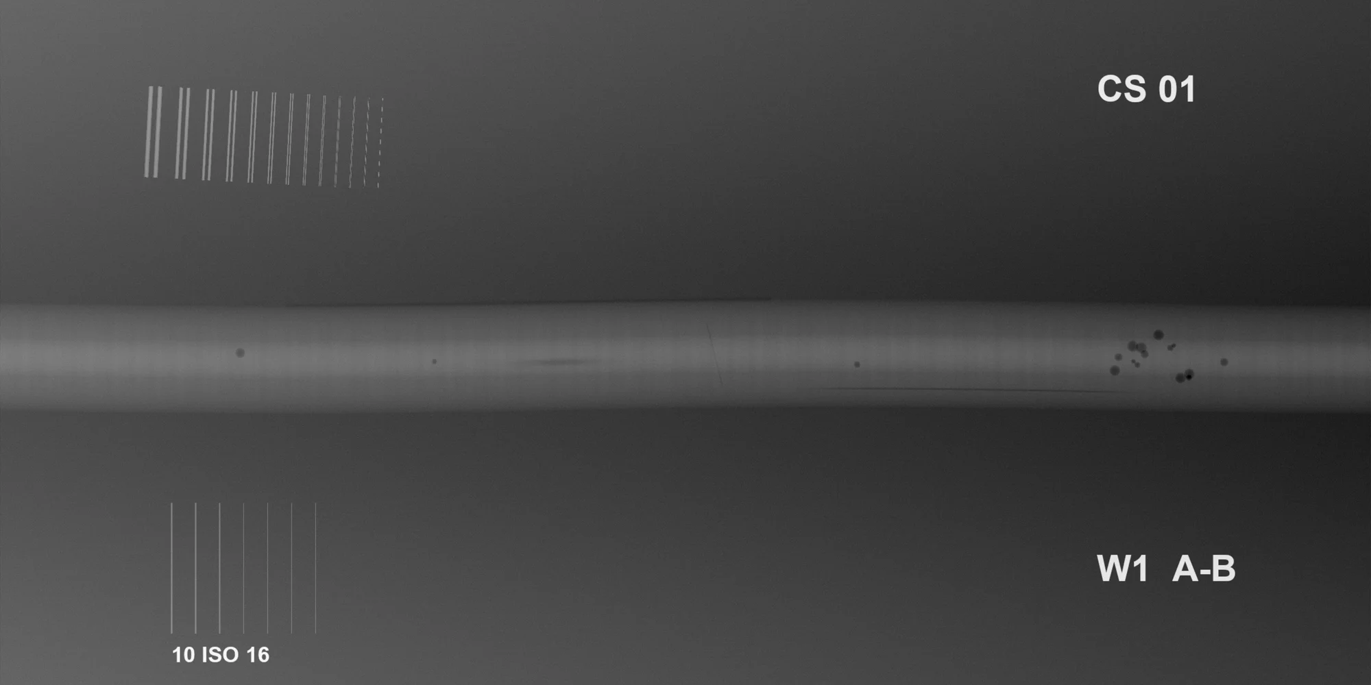
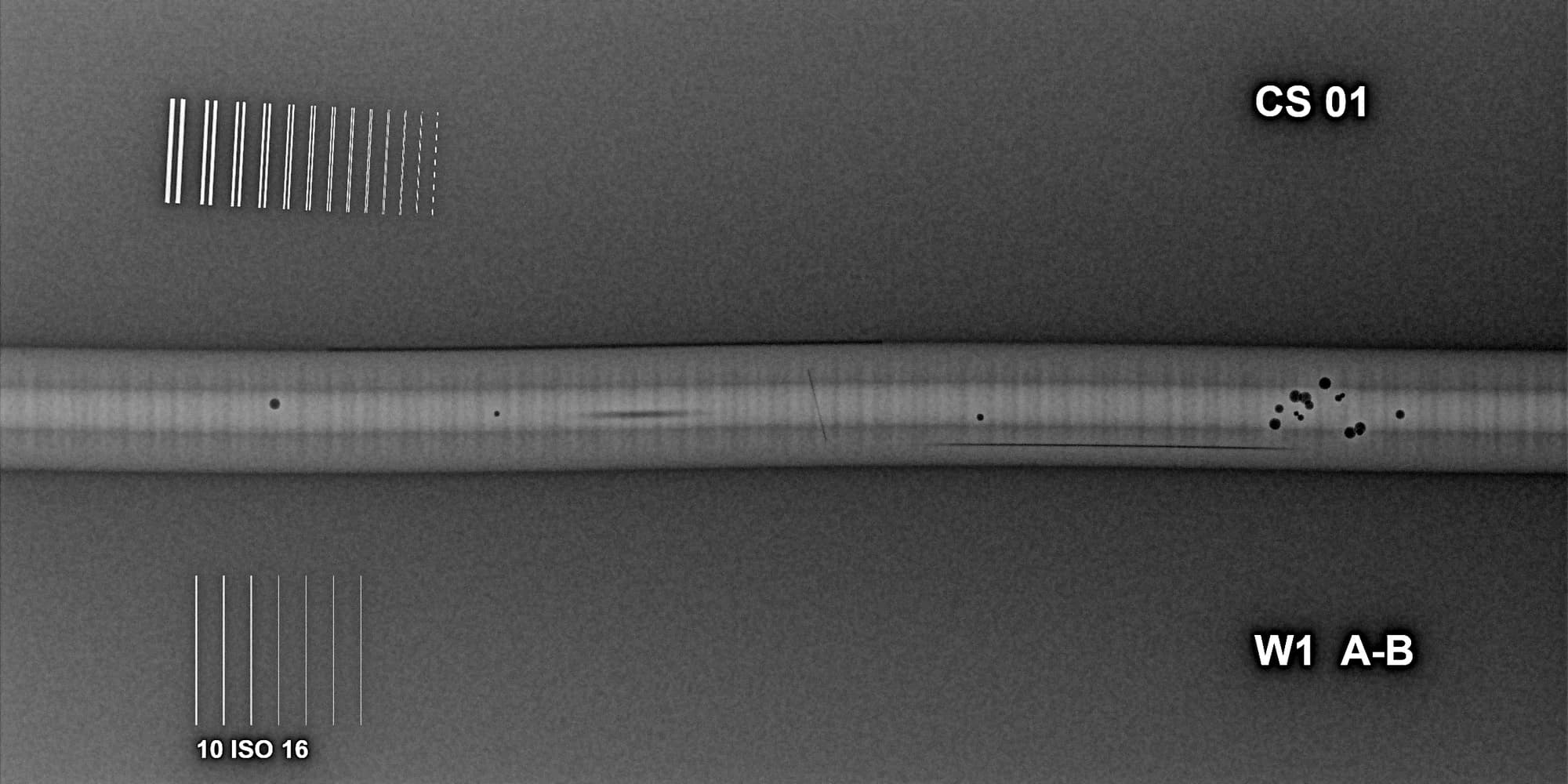
- Normalised SNR
- 89 needs ≥ 70
- ISO 17636-2:2022 Table 3, class A
- Duplex wire IQI
- D9 needs D9
- ISO 17636-2:2022 Table B.13; ISO 19232-5 dip rule
- Pore I1, equivalent diameter
- 1.46 mm
- Sized by region growing; length 1.6 mm
16 ms deconvolution
Ten Richardson–Lucy iterations on a 3056 × 2544 radiograph, on the graphics card. The same job takes 2.7 seconds on the CPU.
mm, not grey levels
Pixel spacing comes from the DICONDE file and thickness from your calibration, so every reading is in millimetres you can put in a report.
ISO 17636-2 built in
Minimum SNRn from Tables 3 and 4, duplex and single-wire requirements from Annex B, read with the ISO 19232-5 dip rule. Cited on every result.
0 installs
It runs in the browser on any PC, tablet or phone. Your clients open shared jobs the same way: no account, just a code sent to their email.
Works with your panel
Opens files from any DR system
Standard DICONDE exports from Carestream, Waygate Technologies (Baker Hughes), Teledyne, Vidisco, Dürr NDT and others open as they are, compressed or not, at their full bit depth. Systems that export TIFF instead work too. Check your system.
- Uncompressed DICOM / DICONDE
- JPEG Lossless
- JPEG-LS
- JPEG 2000
- RLE
- Deflate
- JPEG Baseline
- 8 / 16-bit TIFF
- JPEG / PNG pictures
A lossy-compressed original is flagged on the image, so nobody mistakes it for the full-quality file. JPEG or PNG pictures open too, marked “picture only”. The original you upload is stored byte for byte with its SHA-256 fingerprint.
Every format above is tested on reference files. Product names are trademarks of their owners; Clearshot DR is not affiliated with or endorsed by them.
Everything between the panel and the report
All features in detail- Weld detail preset
Filters and presets
8-band multi-scale enhancement, edge enhance, rolling-ball flattening, FFT de-striping, guided and anisotropic noise reduction, deconvolution. Built-in presets and your own.
- 124 ms, every filter on
WebGPU speed
Heavy filters run on the graphics card. Every filter on a 7.8-megapixel exposure in about an eighth of a second.
- ⌀ 1.46 mm
Calibrated measurement
Length, angle, line profiles and box statistics in millimetres, straight from the pixel spacing in the file.
- 10.92 mm at the defect
Thickness and wall loss
Calibrate on known steps and the image becomes a thickness map. Tangential wall thickness with an uncertainty.
- D9 resolved, needs D9
ISO 17636-2 image quality
SNRn and duplex IQI measured and checked against the 2022 tables for your class and technique, with the table cited.
- I2 ⌀ 0.84 mm
Indication sizing
Outline an indication for length, width, area, equivalent diameter and contrast. Advisory check against the limits you enter.
False colour
Seven colour scales and relief to pull faint density changes out of the grey.
Stitching
Join overlapping exposures of a long seam into one image. Saved as a new file; the originals never change.
Photo overlay
Lay the radiograph over a photo of the actual part from three or four matching points, so the repair team knows where to dig.
AI assist
Suggests possible indications, marker labels and an IQI reading on their own layer. Your interpreter accepts or rejects each one.
- Shared job · expires in 30 days
Enter the code we emailed to j•••@client.com
Open the imagesShare links and PDFs
Branded reports and expiring client links. Clients prove their email with a 6-digit code, then open the same viewer, read-only, free.
- 09:12J. Clientopened the shared job
- 09:14J. Clientdownloaded weld-A.dcm
- 10:02M. Radiographeraccepted AI suggestion I1
- 10:05M. Radiographersaved assessment, 4 filters
- 10:31A. Adminrevoked share link
SHA-256 recorded with every file · nothing is editable
Audit trail
Uploads, assessments, downloads, AI decisions and every share-link open, with who and when.
One exposure. Three ways to read it.
Drag the divider. Switch between weld enhancement, false colour and the calibrated thickness map, all made from the same untouched original.
Drag across the image. Synthetic demo radiograph, generated for this page and processed by the Clearshot engine.
Try the engine
These sliders drive the real Clearshot processing code, running in your browser on a demo weld. Nothing is uploaded and nothing is pre-rendered.
8-band multi-scale enhancement
Edge-preserving guided filter
Window width
Window level
Measure the steel, not the screen
Pixel spacing comes from the file, so every line, box and profile is in real units. Calibrate on known steps and the image becomes a map of the steel.
- Length, angle, profiles and box statistics
- Thickness maps and tangential wall thickness with uncertainty
- Indication sizing, porosity percentage and an advisory rule check
- Stitching and photo overlay; originals untouched
| Indication | Equivalent ⌀ | Length | Contrast |
|---|---|---|---|
| I1 pore | 1.46 mm | 1.6 mm | 7.0 % |
| I2 pore | 0.84 mm | 1.0 mm | 7.1 % |
- Multi-scale enhancement284 ms on CPU, 11 ms on GPU
- De-stripe166 ms on CPU, 15 ms on GPU
- Deconvolution, 10 iterations2,698 ms on CPU, 16 ms on GPU
- Anisotropic diffusion1,109 ms on CPU, 37 ms on GPU
- Guided filter192 ms on CPU, 48 ms on GPU
- Every filter on2,628 ms on CPU, 124 ms on GPU
Measured in Chrome on an Apple-silicon MacBook. Phones and browsers without WebGPU use the CPU path, so results look the same everywhere.
Show it meets the standard
Image quality is read from the radiograph itself and checked against ISO 17636-2:2022 for your class, technique and thickness. The AI only ever suggests; your interpreter decides.
How the standards checks work- Last resolved
- D9
- First unresolved
- D10
- SRb
- 100 µm
- Accepted by M. Radiographer
Rounded indications grouped near the weld centreline
Indication, confidence 82 %
- Pending
Linear indication along the lower fusion face
Indication, confidence 64 %
- Rejected by M. Radiographer
Faint horizontal banding consistent with panel pattern
Artefact, confidence 55 %
Suggestions only. The qualified interpreter records the result; every accept and reject is logged.
Desktop DR software was built for one PC in one bay
| The usual way | With Clearshot DR | |
|---|---|---|
| Viewing | A desktop licence on one PC in one office | Any browser, nothing to install |
| Sending to the client | Screenshots, USB sticks and oversized attachments | One expiring link; every open is logged |
| Image quality | Worked out by hand, when there is time | SNRn and duplex IQI checked against ISO 17636-2, table cited |
| Measuring | A ruler held up to the screen | Calibrated millimetres, thickness, wall loss and sizing |
| Audit | Whatever someone remembered to write down | Exact processing saved with every assessment; originals fingerprinted |
Closing · 55 %
Sealed in the UK. Untouched. Yours.
Radiographs are evidence, and they can carry commercially and security-sensitive detail. Clearshot is built so only the people you invite can see them and every access leaves a record.
Stored in London
Files and records live on Google Cloud in europe-west2. The app servers run in London too.
Encrypted
TLS in transit, Google Cloud encryption at rest.
Originals untouched
Stored byte for byte with a SHA-256 fingerprint you can check.
Separated by company
Every request is checked against your organisation on the server.
Roles and expiring links
Owner, admin, radiographer, viewer and client. Share links expire and can be revoked.
No training by default
Your images never train AI models unless an owner opts in.
Results your client can open
Send a link instead of a USB stick. Clients open the same viewer, read-only, measure for themselves and download the originals. Free for them, logged for you.
- Access
- View, measure, download originals
- Expires
- 30 days by default, up to 90, or when you revoke it
- Cost to your client
- Nothing. No account, just an email code
- Logged
- Every open and download
From £49 a month
Every plan includes every feature. GBP, excluding VAT. 14-day free trial, no card.
Solo
£49 a month
One radiographer
Team
£199 a month
Up to 5 users, shared library, client links
Business
£499 a month from
15+ users, 2 TB, priority support
Questions radiographers ask
Which files does Clearshot DR open?
DICOM and DICONDE files, uncompressed or compressed with JPEG Lossless, JPEG-LS, JPEG 2000, RLE, Deflate or JPEG Baseline, plus 8- and 16-bit greyscale TIFF. Images keep their full bit depth. Lossy-compressed originals (JPEG Baseline, JPEG-LS near-lossless, lossy JPEG 2000) open but are flagged on the image. JPEG, PNG, GIF and BMP pictures open as "picture only": sizes in pixels and no image-quality checks. 12-bit JPEG Extended and HTJ2K are not supported yet. The full list is on the compatibility page.
Which DR systems does it work with?
Any system that exports standard DICONDE or greyscale TIFF. That includes the usual exports from Carestream, Waygate Technologies (Baker Hughes), Teledyne, Vidisco, Dürr NDT and others. Every supported format is tested on reference files, and Carestream exports have been used on real jobs. If a file will not open, the message names its format so you know what to change in your export settings. Product names are trademarks of their owners; Clearshot DR is not affiliated with them. See the compatibility page for each system.
Do I need to install anything?
No. Clearshot runs in a modern browser on Windows, Mac, Linux and tablets. Chrome, Edge and Safari use the graphics card (WebGPU) for the heavy filters; other browsers use the CPU path and give the same result, a little slower.
Is Clearshot a medical device?
No. Clearshot DR is for industrial non-destructive testing only and must not be used for medical diagnosis.
Where is our data stored?
In the United Kingdom, on Google Cloud in London (europe-west2). Each company’s jobs, images and reports are kept separate, encrypted in transit and at rest, and only the people you invite can see them.
Does the AI decide whether a weld passes?
Never. The AI assistant only suggests: possible indications, lead-marker labels, an IQI reading or a preset. Your qualified interpreter accepts or rejects each suggestion and records the result.